Cytoskeleton: Difference between revisions
imported>Anonymous m (Cytoskeleton moved to Cytoskeleton ON WHEELS) |
imported>Larry Sanger m (Cytoskeleton ON WHEELS moved to Cytoskeleton: revert) |
Revision as of 07:30, 15 February 2007
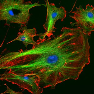
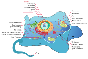
The cytoskeleton is a cellular "scaffolding" or "skeleton" contained, as all other organelles, within the cytoplasm. It is contained in all eukaryotic cells and recent research has shown it can be present in prokaryotic cells too. It is a dynamic structure that maintains cell shape, enables some cell motion (using structures such as flagella and cilia), and plays important roles in both intra-cellular transport (the movement of vesicles and organelles, for example) and cellular division.
The eukaryotic cytoskeleton
Eukaryotic cells contain three kinds of cytoskeletal filaments.
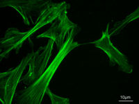
Actin filaments
Main article: actin.
Around 7 nm in diameter, this filament is composed of two actin chains oriented in an helicoidal shape. They are mostly concentrated just beneath the plasma membrane, as they keep cellular shape, form cytoplasmatic protuberances (like pseudopodia and microvilli), and participate in some cell-to-cell or cell-to-matrix junctions and in the transduction of signals. They are also important for cytokinesis and, along with myosin, muscular contraction.
Intermediate filaments
Main article: intermediate filaments
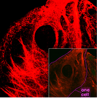
These filaments, 8 to 11 nanometers in diameter, are the more stable (strongly bound) and heterogeneous constituents of the cytoskeleton. They organize the internal tridimensional structure of the cell (they are structural components of the nuclear envelope or the sarcomeres for example). They also participate in some cell-cell and cell-matrix junctions.
Different intermediate filaments are:
- made of vimentins, being the common structural support of many cells.
- made of keratin, found in skin cells, hair and nails.
- neurofilaments of neural cells.
- made of lamin, giving structural support to the nuclear envelope.
Microtubules
Main article: microtubules
They are hollow cylinders of about 25 nm, formed by 13 protofilaments which, in turn, are polymers of alpha and beta tubulin. They have a very dynamic behaviour, binding GTP for polymerization. They are organized by the centrosome.
They play key roles in:
- intracellular transport (associated with dyneins and kinesins they transport organelles like mitochondria or vesicles).
- the axoneme of cilia and flagella.
- the mitotic spindle.
- synthesis of the cell wall in plants.
A fourth eukaryotic cytoskeletal element, microtrabeculae, were proposed by Keith Porter in the 1960s. Porter's lab observed short, filamentous structures of unknown molecular composition in electron micrographs of whole cells. Due to their filamentous appearance and association with known cytoplasmic structures, microtrabeculae were speculated to represent a novel filamentous network distinct from microtubules, filamentous actin, or intermediate filaments. However, they were later shown to be an artifact of certain types of fixation treatments by Hans Ris and others.
The prokaryotic cytoskeleton
The cytoskeleton was previously thought to be a feature only of eukaryotic cells, but homologues to all the major proteins of the eukaryotic cytoskeleton have recently been found in prokaryotes. Although the evolutionary relationships are so distant that they are not obvious from protein sequence comparisons alone, the similarity of their three-dimensional structures provides strong evidence that the eukaryotic and prokaryotic cytoskeletons are truly homologous.
FtsZ
FtsZ, a was the first protein of the prokaryotic cytoskeleton to be identified. Like tubulin, FtsZ forms filaments in the presence of GTP, but these filaments do not group into tubules. During cell division, FtsZ is the first protein to move to the division site, and is essential for recruiting other proteins that produce a new cell wall between the dividing cells.
Scientist's Carl Linane and James Tennyson have recently made major progress while studying these structures and a presentation will be done regarding this in SPHS Discusion Grounds.
MreB and ParM
Prokaryotic actin-like proteins, such as MreB, are involved in the maintenance of cell shape. All non-spherical bacteria have genes encoding actin-like proteins, and these proteins form a helical network beneath the cell membrane that guides the proteins involved in cell wall biosynthesis.
Some plasmids encode a partitioning system that involves an actin-like protein ParM. Filaments of ParM exhibit dynamic instability, and may partition plasmid DNA into the dividing daughter cells by a mechanism analogous to that used by microtubules during eukaryotic mitosis.
Crescentin
The bacterium Caulobacter crescentus contains a third protein, crescentin, that is related to the intermediate filaments of eukaryotic cells. Crescentin is also involved in maintaining cell shape, but the mechanism by which it does this is currently unclear.
Further reading
- Linda A. Amos and W. Gradshaw Amos, Molecules of the Cytoskeletion, Guilford, ISBN 0-89862-404-5, LoC QP552.C96A46 1991
External links
- Cytoskeleton, Cell Motility and Motors - The Virtual Library of Biochemistry and Cell Biology
- Cytoskeleton database, clinical trials, recent literature, lab registry ...
Template:Cytoskeletal Proteins
bn:সাইটোকঙ্কাল cs:Cytoskelet de:Cytoskelett fr:Cytosquelette ko:세포골격 hr:Citoskeleton is:Frymisnet it:Citoscheletro he:שלד התא lb:Zytoskelett lt:Citoskeletas nl:Cytoskelet ja:細胞骨格 pl:Cytoszkielet ru:Цитоскелет sr:Ћелијски скелет sv:Cellskelett zh:细胞骨架


